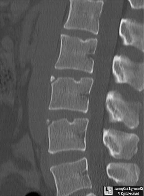
First and quite importantly know that vertebrae and discs dont slip. Cervical intercalary bones and limbus vertebrae have a similar appearance but should not be confused for a cervical spine fracture.

First and quite importantly know that vertebrae and discs dont slip.
Limbus vertebra cervical spine. A limbus vertebra is a well-corticated unfused secondary ossification center usually of the anterosuperior vertebral body corner that occurs secondary to herniation of the nucleus pulposus through the vertebral body endplate beneath the ring apophysis see ossification of the vertebrae. Although Limbus vertebra is not an uncommon radiological finding in an adult it is a rare finding in the child or adolescent. The most common site for the presence of Limbus vertebra is the mid-lumbar region and less commonly occurs in the mid cervical region.
Although Limbus vertebra is not an uncommon radiological finding in an adult it is a rare finding in the child or adolescent. The most common site for the presence of Limbus vertebra is the mid-lumbar region and less commonly occurs in the mid cervical region. Limbus vertebra is a condition characterized by marginal interosseous herniation of the nucleus pulposus and causes non specific symptoms like low back pain back pain muscle spasms and.
The limbus vertebra top shows corner fragments that are well-corticated white arrows The same bodies contain Schmorls nodes yellow arrows. Discitis lower left destroys two adjacent endplates yellow and white arrows and the intervening disk space. The edges are not sclerotic.
Imaging features of posterior limbus vertebrae. 20123606797802 Revista Argentina de Radiología Argentinian Journal of Radiology Vol. 22020 76 Vértebra limbus cervical.
Cervical intercalary bones and limbus vertebrae have a similar appearance but should not be confused for a cervical spine fracture. 2 article feature images from this case 14 public playlist include this case. Limbus vertebra is defined as the presence of an ossicle or an adjacent bone affecting the margin angle of the vertebral bodies.
The characteristic appearance on plain films is. Limbus Bones Limbus bones also known as vertebral edge separations occur primarily in the lumbar spine. Most are identified long after the inciting incident and are asymptomatic.
The radiographic appearance is distinct. A small well-defined and corticated triangular fragment of bone will be present at the anterior superior. A limbus vertebra is a bone tubercle formed by bone trauma on a vertebral body bearing a radiographic similarity to a vertebral fracture.
The anterior-superior corner of a single vertebra is the common site for this defect although it can also be seen at the inferior corner as well as the posterior or anterior margin. Seven cervical vertebrae labeled C1 to C7 form the cervical spine from the base of the skull down to the top of the shoulders. At each level the cervical vertebrae protect the spinal cord and work with muscles tendons ligaments and joints to provide a combination of support structure and flexibility to.
A limbus vertebra is a bone tubercle formed by bone trauma on a vertebral body bearing a radiographic similarity to a vertebral fracture. The anterior-superior corner of a single vertebra is the common site for this defect although it can also be seen at. The cervical spine consists of seven vertebrae and is located at the base of the skull.
Its function is to support the skull enabling head movements back and forth and from side to side as well. Introduction Limbus vertebra LV is a condition characterized by marginal interosseous herniation of nucleus pulposus causing non-specific symptoms such as back pain local muscle spasm and radiculopathy. It is frequently confused with vertebral fracture infection Schmorl nodule or tumor since it does not present any specific symptoms.
Discs sit between the vertebrae in the back and are responsible for absorbing shock and supporting the body. First and quite importantly know that vertebrae and discs dont slip. Its called a slipped vertebra or disc because when it occurs it feels as if something is off in the back.
Images from a 33-year-old female who had low back pain with numbness of the right lower leg for months. A and B noncontrast CT images Posterior limbus vertebra PLV at the superior endplate of the S1 vertebra comprises intravertebral disc herniation through the posterior ring physis white arrows and retropulsion of the posterior ring apophysis limbus fragment black arrows. There are seven cervical vertebrae but eight cervical spinal nerves designated C1 through C7.
These bones are in general small and delicate. Their spinous processes are short with the exception of C2 and C7 which have palpable spinous processes. Smoothly corticated triangular fragment of bone at the corner of the vertebral body Most common in midlumbar spine L2-4 but can be at any level including cervical spine Most common at the anterosuperior corner of the vertebral body Typically displaced farther from margin of vertebral body.
Limbus vertebra was first described by Schmorl in 1927 Schmorl and Junghanns 1971 and results from the herniation of the nucleus pulposus into the body of the adjacent vertebra near its anterior margin causing separation of a triangular bony fragment Fig. A limbus vertebra is a bone tubercle formed by bone trauma on a vertebral body bearing a radiographic similarity to a vertebral fracture. The anterior-superior corner of a single vertebra is the common site for this defect although it can also be seen at the inferior corner as well as the posterior or anterior margin.